
The Human eye is the organ that gives us the sense of sight, allowing us to observe and learn more about the surrounding world than we do with any of the other four senses. Most people probably would agree that sight is the sense they value more than all the rest.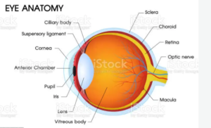
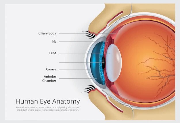
Anatomy of Human Eye:
Before entering with specific parts of the human eye, we should have a gross idea regarding its anatomy-
Sclera
The sclera is the tight white sheath that forms the outer layer of the ball. It is also referred to by other terms, including the sclerotic and the sclerotic coat( both having precisely the same meaning as the sclera)
Functions:
- Protect its delicate internal parts
- Meet the purpose of attachment of the extraocular muscles that enable eyes to move.
- Maintains the shape of the eye
Disease of sclera
Scleritis is a disease of sclera but not a common one. It is inflammation of that white part of the eye. Sometimes sclera gets hurt and this serious condition occurs.
Causes of scleritis
There is no certain reason for this disease. Eye injury, serious allergy, or infection can cause scleritis. Again experts say that it has associations with some other diseases also. Episcleritis may convert into scleritis. The inflammation of the episclera is ‘Episcleritis’. The diseases are-
- Lupus
- Systemic sclerosis or Scleroderma
- Sjogren’s syndrome
- Different types of inflammatory arthritis. For example- Reactive arthritis.
- Inflammatory bowel disease
- Rheumatoid arthritis etc.
Symptoms
There are two types of scleritis and the signs are also different.
Anterior: Around 98% of the cases this type occurs. It causes inflammation in the front part of the sclera.
Posterior: It causes inflammation in the back part of the sclera. This uncommon type may cause serious eye problems like glaucoma.
Treatments
The treatments depend on the condition of the eyes. Ophthalmologists suggest medicines to relieve pain. They suggest surgeries in serious cases. Again, corneal glue, bandage contact lenses to repair damages of corneal tissues.
Conjunctiva
The conjunctiva is a transparent mucous membrane consisting of cell and underlying basement membrane that covers the sclera( white part of the eye) and lines the inside of the eyelids.
Functions:
- It helps lubricate the eye by producing mucus and tears, although a smaller volume of damages than the lacrimal gland.
- It also contributes to immune surveillance and helps to prevent the entrance of microbes into the eye.
Diseases of conjunctiva
Conjunctivitis
Sometimes viruses and bacteria can grow in the moist environment of the conjunctiva. Inflammation occurs and causes discomfort. Allergies, injuries, chemical reactions can also cause conjunctivitis.
Youngs is the victim of vernal conjunctivitis often. This happens in summer and spring.
Signs
- Red eyes
- Pink eyes
- Thick discharges
- Internal or external Irritation
- Swollen eyelids
- Pain
Treatments
Antibiotic eye drops, medicines of allergy, and anti-inflammatory can solve these conditions. Doctors also prescribe surgeries and steroids. But, some serious conjunctival disorders can cause sight loss. So, we should not ignore eye problems and consult our eye specialists.
Pupil
The pupil is the hole in the center of the iris that light passes through the iris muscles to control its size.
Functions:
- Regulates the amount of light passing through to the retina
- Acts as an aperture of a camera
- When dilated by mydriatic agent, it causes the beautification of eyes followed by the ancient ladies.
Disorders of pupil
Sometimes injuries cause problems in pupils. Sizes of pupils matter but injuries due to surgeries may change the shapes. Disorders of pupils depend on several facts include-
Coloboma-
Before birth, some children do not get the right shapes of eyes. Children then suffer from this disease after birth. Irish with coloboma makes the pupils very big. And we notice the change in shapes.
Anisocoria–
In this case, one size of the pupil is bigger than the other one. It can happen all of a sudden and without reason. Sometimes people feel abnormalities also. So, we should consult doctors.
Horner’s Syndrome-
The nerves of eyes are very sensitive. Any problem or injury in nerves may lead to this syndrome. Again, sometimes, children take birth with this disorder. It makes a pupil small in shape.
Brain and Headache Problem-
Tumor in the brain, head injuries, headache can cause disorder of pupils. Again, a brain stroke is also a reason for it.
Adie Syndrome-
Proper blood flow is necessary into the eye nerves. Lack of it can lead to this syndrome. An injury is also a cause but there is no certain reason for it. People with this problem can’t react to light properly.
Treatments
Proper medicines and suggestions of specialists can solve these disorders. Medicines include motion sickness medicines, atropine, antihistamines, etc. are essential.
Cornea
The cornea is the transparent circular part in the front of the human eyeball. It has an essential optical function as it refracts light entering the eye through the pupil and onto the lens.
Functions:
- It refracts the light entering the eye through the pupil and onto the lens.
- Protects the pupil and iris from the external environment.
Corneal diseases
People suffer from many inflammations of the cornea. Some diseases are-
- Corneal Ulcer
- Herpes Zoster Ophthalmicus
- Dystrophies
- Trauma
- Herpes Simplex Keratitis
- Nutritional deficiencies
- Keratoconus etc.
Signs of corneal diseases
- Sensitivity of light
- Cloudy sight
- Fatigue
- Pain
- Tearing
- Headache
- Nausea
Treatments
The diseases are different. So, treatments depend on patients’ conditions. Eye doctors need to check and test them. Some treatments are-
- Medicines
- Eye drops
- Ointments
- Oral medication
- Surgery
- Laser treatment etc.
Iris
The iris is the colored part of the eye. It is a thin diaphragm composed mostly of connective tissue and smooth muscle fibers. The iris lies between the cornea and the crystalline lens.
Functions :
- Gives our eyes a particular color example blue, green and brown
- Responsible for controlling the diameter and size of the pupils
- Responsible for controlling the amount of light reaching the pupil.
Diseases of iris
There are many types of iritis. It is the inflammation of iris. It sometimes attacks the uvea are then we call it uveitis. These problems sometimes become very serious. Existing health problems, eye injuries, side effects of medicines lead to these sicknesses.
Symptoms of iritis
- Red eyes
- Different abnormal reactions in light
- Blurry vision
- Headache
- Problems in the shapes of pupils
- Pain and irritation in eyes
- Loss of vision
Treatments
These eye complications need medical care no doubt. Eye specialists do blood tests, x-ray and many other tests to diagnose the actual problem. Then they suggest medications to the patients.
Dysfunctions of the ciliary muscle
Many eye sicknesses have involvement with these muscles. Researchers say that people with Adie’s syndrome sometimes have ciliary muscle dysfunctions. But proper medications can solve these problems.
Lens
The lens is a transparent structure enclosed in a thin transparent capsule. It is located behind the pupil of the eye and encircled by the ciliary processes.
Functions :
- Producing an image of the object on to the retina
- Acts like the lens of a camera
- The lens of the eye helps to refract light traveling through the eye ( which first refracted by the cornea) the lens focuses light into an image on the retina. It can do this because the shape of the lens is changed according to the distance from the eye of the objects the person is looking at
This adjustment of the shape of the lens is called accommodation and is achieved by the contraction and relaxation of the ciliary muscle.
Problems in lens
The lens is an important part of human eye anatomy. People face many eye lens complications include age-related problems. For examples-
- Two types of cataract: Congenital and Acquired
- Congenital: coloboma, lentiglobus, mittendorf dot, etc.
- Microspherophakia
- Parallax
- Ectopia lentis
- Nance-Horan syndrome etc.
Signs
- Slow diminution of sight without pain
- Black spot in the field of vision
- Low sight
- Headache
- Polyopia problem
- Glaucoma
Treatments
These patients need urgent consultation with doctors. Doctors can prescribe medications depending on the conditions.
Ciliary muscle
Contraction and relaxation of the ciliary muscle alters the curvature of the lens.
Functions :
- Helps to alt the curvature of the lens
- Changes focal length of the lens
Dysfunctions of the ciliary muscle
Many eye sicknesses have involvement with these muscles. Researchers say that people with Adie’s syndrome sometimes have ciliary muscle dysfunctions. But proper medications can solve these problems.
Choroid
The choroid is the layer of the eyeball located between the retina and sclera. It is a thin, highly vascular( i.e., it contains blood vessels) membrane.
Functions :
- It supplies blood to the retina.
- contains a pigment that absorbs excess light and so prevents blurred vision (due to too much light on the retina).
- It provides oxygen and nourishment to the outer layers of the retina.
Problems of Choroid
There are many names of these sicknesses of eyes. Some are-
- Different kinds of choroidal dystrophies
- Choroidal folds
- Neoplasm
- Detachment and hemorrhage of the choroid
- Choroidopathy
- Abnormalities of the vascular layer
- Choroidal rupture etc.
Symptoms
Some kinds of diseases are results of abnormal genes. These occur in childhood. But, some other types occur later. Examples of sicknesses are- pain, vision loss, dysfunctions of the retina, etc.
Treatments
Some diseases do not need any treatment and some of them have none. But, still, there are medications of some complications include-
- Systemic steroids
- Laser treatments
- Eye pressure-lowering drugs
- Surgery etc.
How to Maintain Good Eyesight?
Twelve foods to boost your vision are-
- Nuts
- Legumes
- Carrots
- Avocados
- Leafy greens
- Kale
- Sweet potatoes
- Eggs
- Broccoli
- Salmon
- Tuna
- Sunflower seeds
Five good rules to follow are-
- Proper sleep
- A correct weight
- Wear necessary glass
- Regular exercise
- Know the medical history of your gene